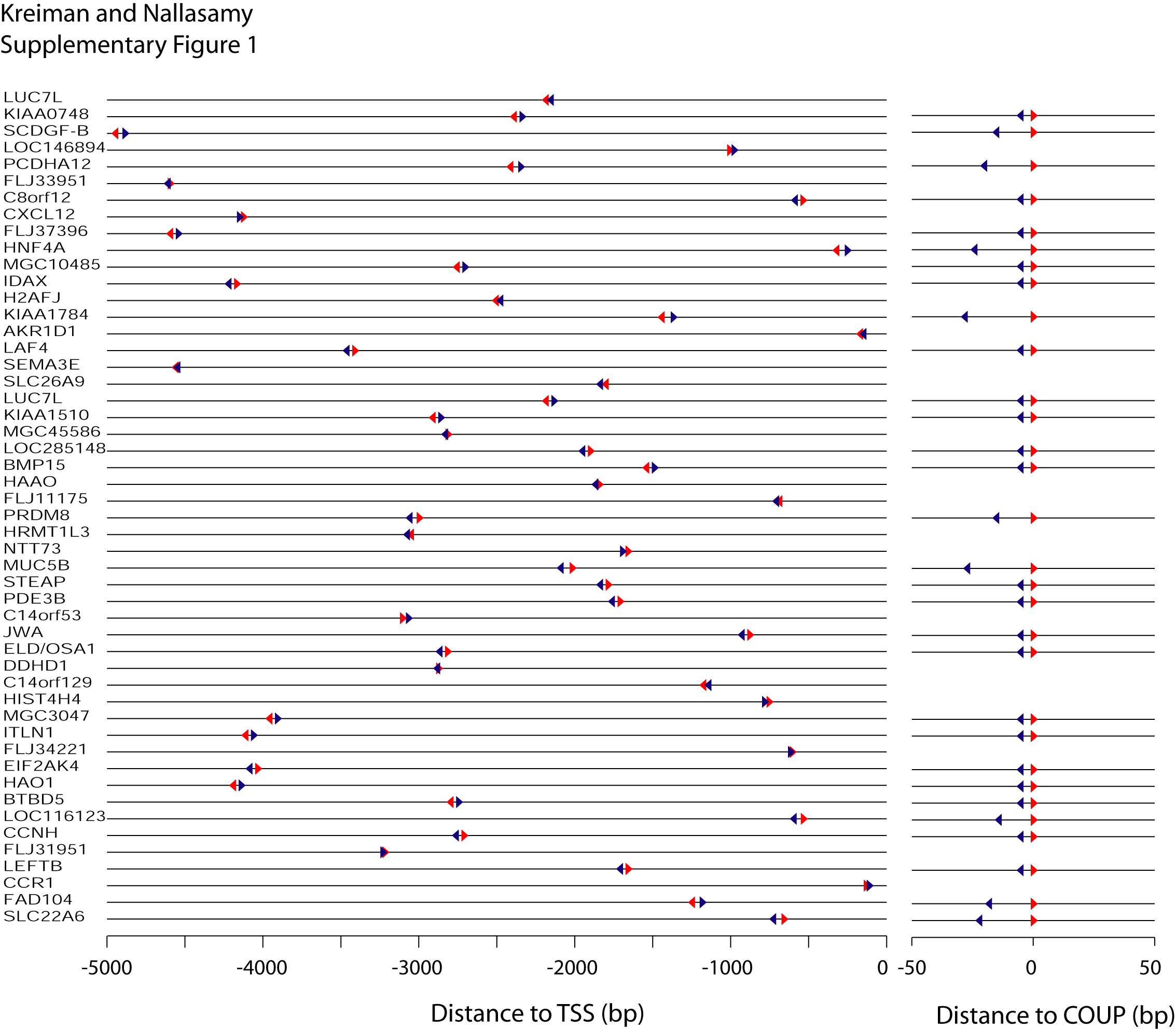
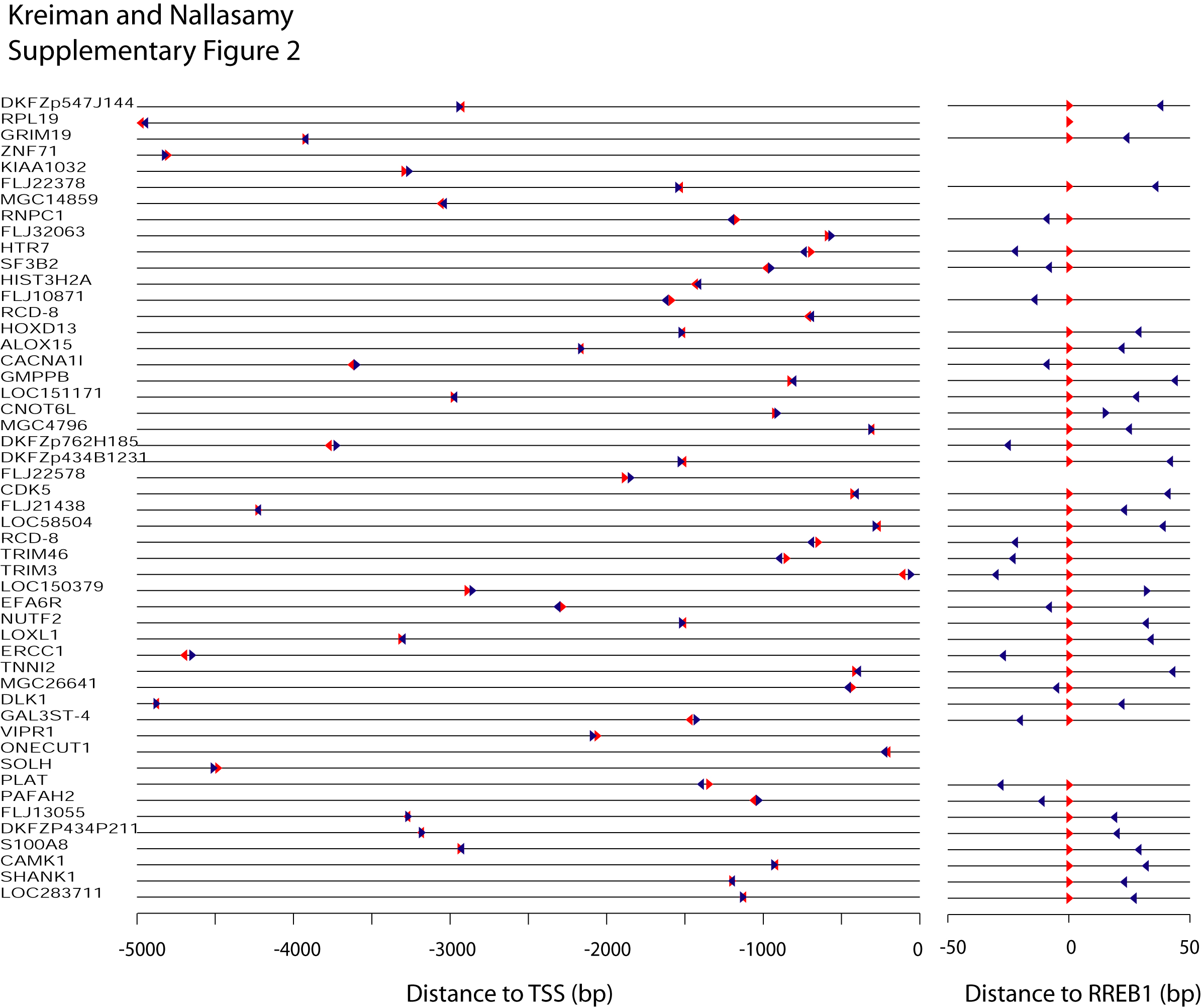
Supplementary Figure 2
Click on Figure to enlarge
Example of two PWMs (RREB1 and P300) where there was a bias towards co-occurrence
on opposite strands (patterns C and D).
(A) Positions of RREB1 (red)
and P300 (blue) putative binding
sites in 50 randomly selected genes where the two motifs co-occurred within
50 bp.
The format and conventions follow the ones in Figure 2 in the main text. Positions
are given with respect to the annotated transcription start site of each gene
(TSS).
Leftward pointing triangles indicate occurrences in the minus strand and rightward
pointing triangles indicate occurrences in the plus strand.
(B) Zoom-in (10x) showing binding sites aligned to the putative
binding sites of RREB1 (red) for
those cases in which RREB1 and P300 co-occurred in opposite strands.