
Figure
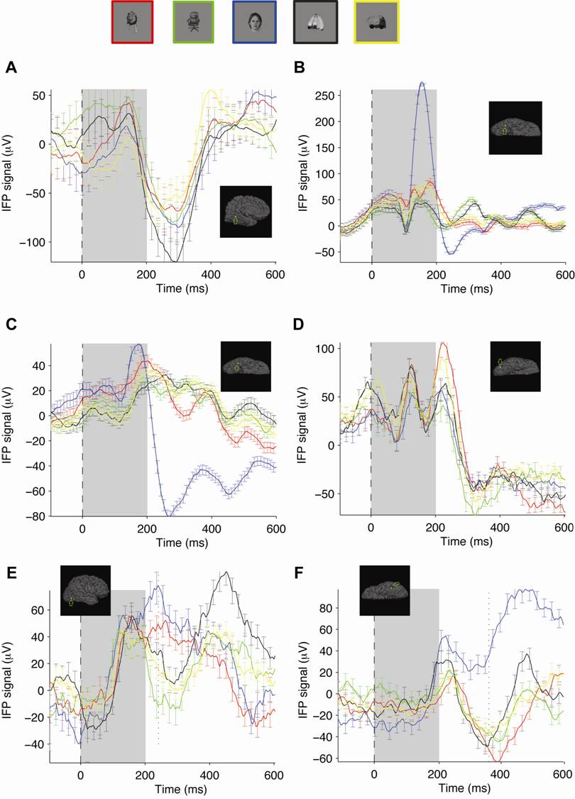
S3: Multiple examples showing selectivity in IFP recordings
Examples
from six electrodes that showed visual selectivity. The format and conventions are
the same as in Figure 1A.
The gray rectangle indicates the image presentation time and the electrode
positions are indicated by the arrows in the small insets. Electrode locations:
(A) left medial temporal (Talairach
coordinates: -12.1 -58.3 -7); (B, C)
right posterior subtemporal (Talairach coordinates: 43.4 -51.3 -13.1 and 48.3
-44.5 -19.3); (D, E)
right posterior temporal (Talairach coordinates: 27.0 -66.0 3.0 and 38.0 -62.0
15.0); (F) left lateral temporal (Talairach
coordinates: -39.7 -42.8 -20.0).