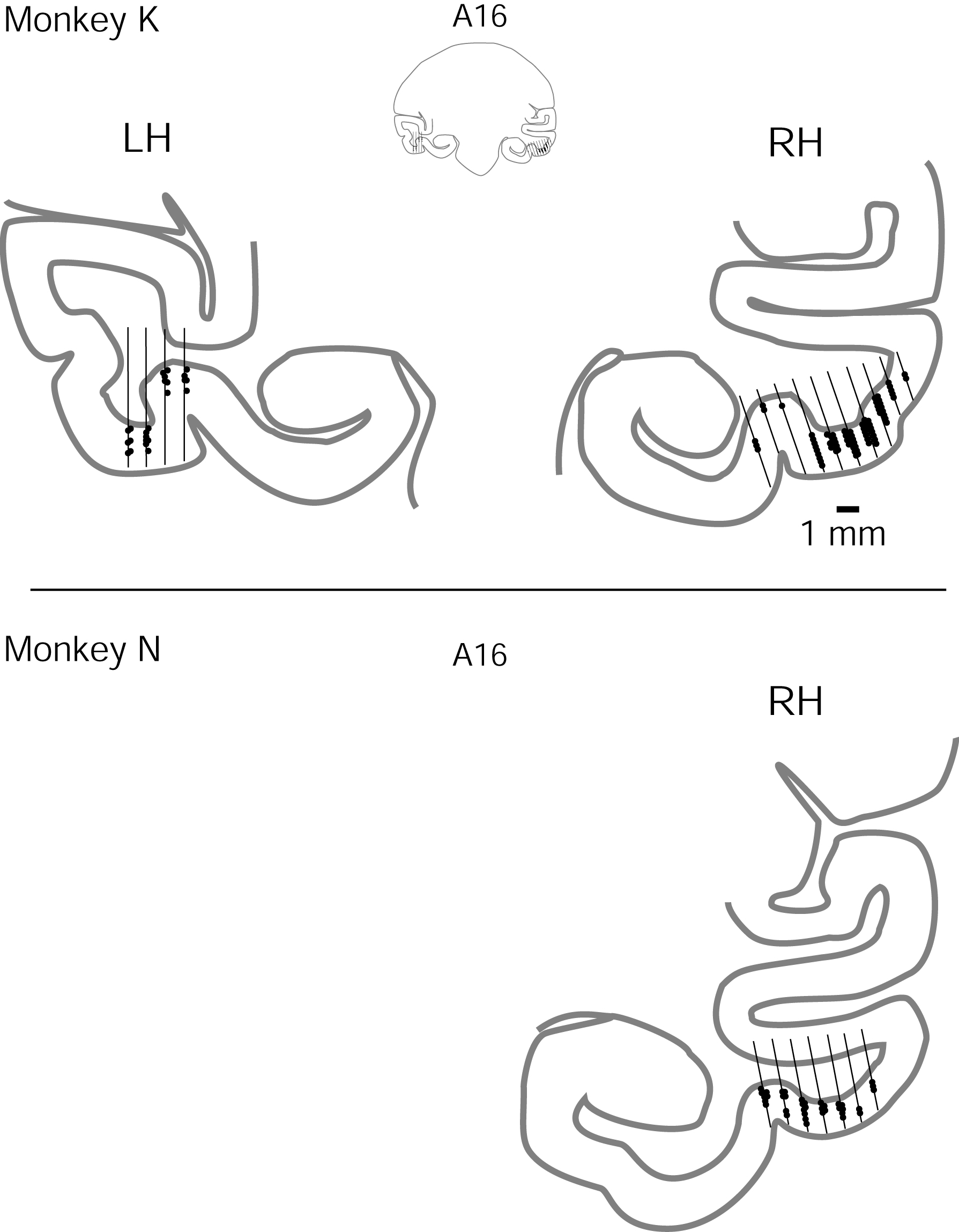
Estimates of electrode penetrations (lines) and recording site locations (dots) are drawn based on MR images from both monkeys taken after chamber implantation. Electrode penetrations were spaced 1 mm apart using a fixed grid at the surface of the skull and true tracking of each penetration was assisted by a guide tube that extended to within 20 mm of the ventral surface. The coronal sections at A16 are shown with recording sites for A15–A17. Only the right hemisphere was recorded in monkey N. Map topography was confirmed by the audible transitions between gray and white matter and by the depth of electrode travel before reaching the ventral surface and (in some cases) the dura beyond the ventral surface. Recording sites were estimated on this plot based on the point of physiologically assessed entry in the gray matter of the ventral surface. Note that electrode tracks were generally aligned along the columnar axis, and fewer recordings were made where alignment was poor.