Biological and Computer Vision
Gabriel Kreiman
Cambridge University Press. 2021. ISBN 9781108649995
Additional Materials
Chapter V: Adventures into terra incognita: probing the neurophysiological responses along the ventral visual stream
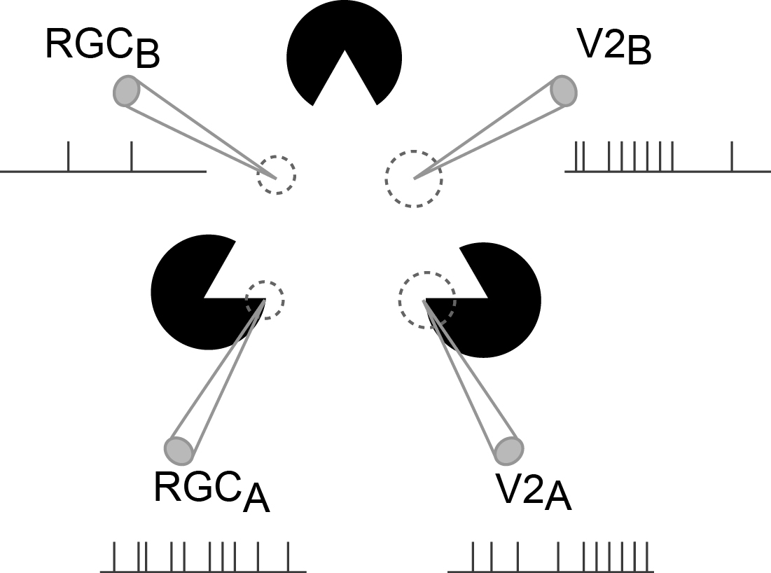
Heroic efforts have begun to elucidate how neurons represent information along the visual cortex. Cortex is characterized by a stereotypical connectivity pattern, a canonical microcircuit that is repeated over and over again in an approximately hierarchical arrangement. The gold standard to examine neural computations is to record the activity of individual neurons. Neurons within each visual area have localized receptive fields that jointly map the entire visual field. Hubel and Wiesel discovered that neurons in the primary visual cortex (V1) selectively respond to oriented bars, thus accentuating the edges in an image. A first-order model suggests how the tuning of neurons in the primary visual cortex can be explained by pooling the activity of adequately aligned input neurons from the lateral geniculate nucleus with circular center-surround receptive fields. Hints of how neurons explicitly represent an interpretation of the visual world rather than precisely the sensory inputs are given by the responses to illusory contours.